Introduction
Skin is the largest organ of the body with a surface area of 2 m2 in a 70 kg individual. It accounts for 16 to 20% of the total body weight. Human skin is of two types: hair-bearing skin and non-hair-bearing (glabrous) skin as seen on palms and soles. The glabrous skin is marked by a series of ridges and grooves (sulci) with a configuration unique to each individual known as dermatoglyphics. Skin is primarily composed of the following: 1. Epithelial tissue (epidermis and dermis) 2. Adipose tissue (hypodermis)
3. Accessory structures (hair, nails, glands and sensory receptors).
Structure of the Skin

The skin is composed of three layers:
- The epidermis: contains keratinocytes which synthesize keratin (protective protein)
- The dermis: made up of fibrillar structural protein known as collagen.
- Subcutaneous tissue (hypodermis/panniculus) : contains lipocytes
Hair, nails, sebaceous, sweat and apocrine glands are regarded as derivatives of skin.
EPIDERMIS
Epidermis: It comprises of stratified squamous epithelium, which is 0.4 to 1.5 mm thick whereas the entire skin is up to 4 mm in thickness. It is composed of five layers.
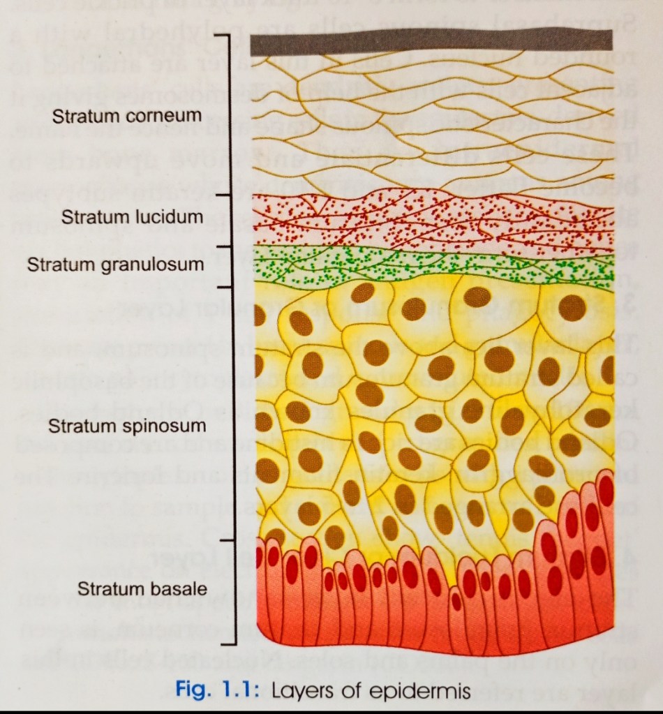
1. Stratum Basale or Stratum Germinativum: It is a single cell layer but may become 2-3 cells thick in hyperproliferative epidermis and glabrous skin. Morphologically, the cells of this layer are small and cuboidal (10-14 um in diameter) with dense cytoplasm which contains tonofilament bundles, ribosomes andi large, dark-staining nuclei. The cells are attached to each other through desmosomes and to the basement membrane zone (BMZ) with the help of hemidesmo somes via keratin filaments K5 and K14. Cell division at this level occurs every 18th to 19th day making it the primary site for mitotically active cells. Melanocytes are seen interspersed in the basal layer. Basal layer mainly consists of three types of cells—stem cells, transient amplifying cells and postmitotic cells.
2. Stratum Spinosum or Prickle Cell Layer: Stratum spinosum is immediately above the basal cell layer, wherein the basal keratinocytes increase in size and number to form 8-10 thick laver of prickle cells. Suprabasal spinous cells are polyhedral with a rounded nucleus. Cells in this layer are attached to adjacent cells with the help of desmosomes giving it the characteristic spinous shape and hence the name. These cells differentiate and move upwards to become flatter. K1 and K10 are keratin subtypes abundantly seen. Stratum basale and spinosum together form the malpighian layer.
3. Stratum Granulosum or Granular Layer: This layer lies above the stratum spinosum, and is called stratum granulosum because of the basophilic keratohyaline granules known as Odland bodies. Odland bodies are rich in histidine and are composed of profilaggrin, keratin filaments and loricrin. The cells are arranged in 2 to 5 layers.
4. Stratum Lucidum or Clear Cell Layer: The electron lucent layer sandwiched between stratum granulosum and stratum corneum, is seen only on the palms and soles. Nucleated cells in this layer are referred to as transitional cells.
5. Stratum Corneum or Dead Cell Layer: It is the outermost skin layer with thickness of 20-25 cells. Cells in this layer are known as corneocytes which are anucleate, flattened cornified cells that are a result of complete differentiation of granular cells. The cells that were originally attached by desmosomes, separate to become flattened; and are shed while moving towards the surface. The main function of this layer is to provide mechanical protection to the skin and act as a barrier to water loss and permeability of soluble substances from the environment.
Types of Cell in Epidermis
- Keratinocytes: (90% of the cells) produce keratin which is a tough fibrous protein that provides protection.
- Melanocytes: which produce the pigment melanin that protects against damage by ultraviolet radiation.
- Langerhans cells: involved in immune responses, arise from red bone marrow.
- Merkel cells: which function in the sensation of touch along with the adjacent tactile discs
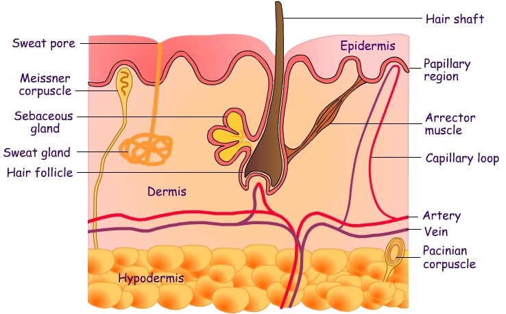
DERMIS
It constitutes 15-20% of the total weight of human body. Dermal thickness may vary depending on the site, e.g. 5 mm over the back and 1 mm over the evelids. It is a complex of fibrous, filamentous and amorphous connective tissue that consists of nerves, vessels, skin appendages, fibroblasts, macrophages, mast cells, Iymphocytes, plasma cells and other leukocytes. Fibrous connective tissue includes collagen and elastic fibers. Non-fibrous connective tissue consists of fine filamentous glycoproteins, proteoglycans (PGs) and glycosaminoglycans (GAGs) of the ground substance. It forms the bulk of skin and provides flexibility, stretch and tensile strength.
Dermis is divided into papillary and reticular dermis. Subpapillary plexus of blood vessels is present between papillary and reticular dermis. Adventitial dermis comprises of papillary dermis and periadenexal dermis.
Dermis Contains Two layers
- Papillary Region (upper layer immediately beneath epidermis) consists of areolar connective tissue containing thin collagen and elastic fibers, dermal papillae (including capillary loops), corpuscles of touch and free nerve endings. It is superficial and thin part of dermis (1/10th) that is present on under surface of the epidermis and forms ridges. It consists of bundles of collagen fibrils (small) and oxytalan elastic fibers. It shows high fibroblastic activity, high metabolic rate and svnthesizes different PGS.
- Reticular Region the deep layer, consists of dense irregular connective tissue containing collagen and elastic fibers adipose cells, hair follicles, nerves, sebaceous (oil) glands, and sudoriferous (sweat) glands. It comprises the lower 9/10th of the dermis and merges with the subcutaneous fat. Reticular dermis is subdivided into upper zone (intermediate size collagen fiber bundles and elaunin fibers) and deeper zone composed of large collagen fibrils.
The Cell of Dermis
- Fibroblasts: are derived from mesenchymal tissue. They are the most common cells found in the dermis. They are seen as bipolar spindle cells with ovoid nucleus. Fibroblasts cause production and degradation of fibrous and non-fibrous proteins of connective tissue and matrix and hence act as a source of ground substance. They also provide a structural framework and help in interactions between epidermis and dermis.
- Monocytes, Macrophages and Dendrocytes: Monocytes, macrophages and dendrocytes together form a mononuclear phagocytic system. Monocytes differentiate into macrophages. They contain lysosomes and phagocytic vacuoles. Dendrocytes are stellate, dendritic or spindle-shaped cells. These cells are phagocytic.
- Mast Cells: Ovoid or spindle-shaped cells seen in the dermis, mast cells are derived from pleuripotent cells in the bone marrow. Mast cells have secretory properties. They are oval-shaped with round nucleus, 6-12 um in diameter and resemble a fried egg. Mast cells are of two types, those found in the dermis and submucosa (type 1) ); and those found in the bowel and respiratory mucosa (type 2). They have a high content of heparin, histamine, neutrophil, eosinophil chemotactic factors, tryptase, kininogenase and B-glycosaminidase. These cells consist of twc components: lamellae (thick, curve, parallel filaments forming whorls) and fine granular material.
Function of Skin
- Regulation of body temperature.
- Blood reservoir.
- Protection.
- Cutaneous sensations.
- Excretion and absorption.
- Synthesis of vitamin D.
Thanks you!
